From Smear to Diagnosis: Simplifying Veterinary Cytology
November 4, 2026
For many veterinary students and early-career doctors, cytology can feel a bit intimidating. Staring down a microscope at clusters of cells, granules, and potential malignancies can make anyone second-guess their diagnostic skills.
To take the mystery out of the process, Vetcor Vice President of Veterinary Leadership, Dr. John Calhoun, recently hosted an exclusive webinar for veterinary students, "From Smear to Diagnosis: Simplifying Veterinary Cytology.” Dr. Calhoun broke down slide interpretation into an approachable, systematic flowchart, proving that with the right mindset and a simple needle and syringe, you can get rapid answers right in the clinic. Here is a recap of the key takeaways to help you master the art of cytology:
The Golden Rule: Don't Be Afraid to Try Again
Cytology is a safe, low-risk, and cost-effective procedure. If your first slide only yields blood or isn't diagnostic, don't hesitate to take a quick breather, bring the patient back, and try a second aspiration. Two slides from the same patient can look completely different, and persistence often pays off with the right answer.
Master the Core Diagnostic Flowchart
Dr. Calhoun emphasized that every mass and fluid sample becomes much easier to evaluate if you follow a simple, step-by-step mental flowchart:
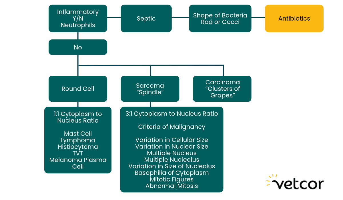
Step 1: Is it inflammatory or non-inflammatory?
Look for neutrophils and macrophages.
- Pro-tip: Be cautious when evaluating severe inflammation or spun fluid samples (like thoracic or abdominal taps), as normal cells like fibroblasts, mesothelial cells, and "fluffy" macrophages can sometimes look deceptively wild and angry.
Step 2: Is it septic or non-septic?
Check for bacteria within the neutrophils. Identifying whether you are dealing with cocci or rods right away gives you an immediate head start on choosing the right antimicrobial therapy.
Step 3: If non-inflammatory, categorize the tumor type.
If the sample is non-inflammatory, your next goal is to place the cells into one of three major buckets:
-
Round Cell Tumors: Characterized by a 1:1 cytoplasmic-to-nuclear ratio and distinct, individual cells. (Think histiocytomas, lymphoma, plasma cell tumors, mast cell tumors, TVTs, and melanomas).
-
Sarcomas (Spindles): Shaped like footballs or "flames," these connective tissue tumors tend to exfoliate separately and behave like an octopus—locally invasive with tentacles that require wide surgical margins.
-
Carcinomas (Glandular Clusters): Glandular cells that typically exfoliate well in "clusters of grapes" and have a higher tendency to metastasize via lymphatics or the bloodstream.
Step 4: Check for criteria of malignancy.
Look for variation in cell and nuclear size, multinucleation, abnormal mitotic figures, and, most importantly, prominent or multiple nucleoli, which signal that cells are reproducing abnormally fast.
Pearl of Wisdom: Always Use Your Microscope!
As one of Dr. Calhoun's former professors noted: "You can't make a microscopic diagnosis without the aid of a microscope." Never bet on physical touch alone. For example, clinicians often assume a subcutaneous mass is a boring, harmless lipoma, only to be surprised by a sneaky mast cell tumor under the lens. Cytology is fast, affordable, and protects you and your team from surgical surprises down the road.
Ready to Grow Your Skills?
Mastering practical skills like cytology doesn't happen overnight, but you don't have to navigate it alone. Whether you're heading into clinical rotations or preparing for your first role in general practice, having a supportive, collaborative team behind you makes all the difference. Ready to start your veterinary journey with a team that has your back? Explore our student programs and open positions today!